Why a cow eye dissection? In large part, because of its similarities to the human eye.
The human eye is one of the most complex and sophisticated organs in the body. Although it’s small and delicate, the eye allows us to see the world without any conscious effort. For example, it adjusts to light automatically. This process enables us to see in both starlight and the brightest sunlight.
The eye’s automatic focusing system is faster and more precise than that of any camera. While a camera lens must be moved back and forth to adjust for distance, the lens of the human eye simply changes shape. (Without this feature, like cameras, we’d need long tubes sticking out of our eyes!)
Each fragile part of the eye works together to provide information to the brain, and the brain interprets it instantaneously giving you a perfect image. It is an amazing process.
Download: Cow Eye Dissection Lab
A cow’s eye, like other farm animal organs, is similar to our eyes. One benefit of a cow eye dissection is that by examining the anatomy of a preserved eye, you can learn how your own eye forms images of the world and sends them to your brain. Another is career potential: farmers, ranchers, and veterinarians (among others) must be intimately familiar with cow eyes to perform their jobs well.
This cow eye dissection kit comes with everything you need to conduct a lab examination.
Cow Eye Observation: External Anatomy
 |
| Click image for full-size pdf |
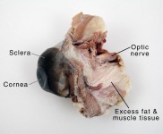
Look carefully at the preserved cow eye. The most noticeable part of the eye is the large mass of gray tissue that surrounds the posterior (back) of the eye and is attached to the sclera. The second most noticeable part of the eye is the cornea, located in the anterior (front) part of the eye. Due to the fact that the eye has been preserved, the cornea is cloudy and bluish-gray in color. It may also be wrinkly and seem a bit ‘deflated’. On the posterior side of the eye, nestled in the fat and muscle tissue, there is a noticeably round protuberance that feels stiffer than the surrounding tissue. This is the optic nerve, and it sends the images collected in the eye to the brain.
Cow Eye Dissection: Internal Anatomy
1. Place the cow’s eye on a dissecting tray. The eye most likely has a thick covering of fat and muscle tissue. Carefully cut away the fat and the muscle. As you get closer to the actual eyeball, you may notice muscles that are attached directly to the sclera and along the optic nerve. These are the extrinsic muscles that allow a cow to move its eye up and down and from side to side. Keep cutting close to the sclera, separating the membrane that attaches the muscle to it. After removing the excess tissue, the sclera and optic nerve should be exposed but still intact.
 |
| Click image for full size pdf |
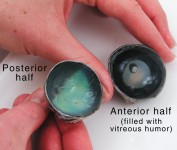
2. Using a sharp scalpel, cut through the sclera around the middle of the eye so that one half will have the anterior features of the eye (the cornea, lens, iris, and ciliary body) and the other half will contain the posterior features (most noticeably where the optic nerve is attached to the eye). The inside of the eye cavity is filled with liquid. This is vitreous humor. Depending on how the specimen was preserved, it will be either a dark liquid that will flow out easily or a slightly gelatinous material that you can pour out to remove. (In a living eye, the vitreous humor is clear and gel-like.)
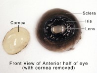
3. Flip the anterior half of the eye over so that the front of it is facing upward. Using a pair of sharp scissors, cut the cornea from the eye along the boundary where the cornea meets the sclera. When the scissors have cut in far enough, clear fluid will start to seep out – this is the aqueous humor. While cutting out the cornea, be careful to not accidentally cut the iris or the lens. After removing the cornea, pick it up and look through it. Although it is cloudy due to the degrading of the tissue, it is still fairly transparent. Notice the toughness and strength of the cornea. It is designed this way to protect the more delicate features found inside the eye.
 |
| Click image for full size pdf |
4. With the front of the anterior half of the eye facing up, locate the iris. Notice how the iris is positioned so that it surrounds and overlaps the lens. This position allows the iris to open and close around the lens to allow different amounts of light into the eye. In bright light, the iris contracts to let in less light. In dim light, such as at night, the iris expands to let in more light.
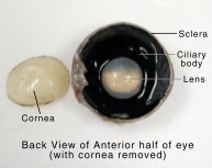
5. Flip the anterior half over and examine the back half. Locate the lens and ciliary body. The ciliary body surrounds the lens, allowing it to change the shape of the lens to help the eye focus on the object it is viewing.
6. After examining both sides of the anterior half of the eye, pull the lens out. While the cow was alive, the lens was clear and very flexible. In a preserved cow eye, the lens will most likely have yellowed and become very hard. However, it may still be possible to look through the lens and see its ability to magnify objects. Try this by placing the lens on a piece of paper with writing on it.
 |
| Click image for full size pdf |
7. On the posterior half of the eye, there is a thin, tissue-like material that slides easily inside the sclera. This is the retina. The retina contains photoreceptor cells that collect the light entering the eye through the lens from the outside world. These images are sent to the optic disc, the spot where the optic nerve attaches to the eye.
At this point, there are no photoreceptor cells; there are only nerves sending images to the brain. Because of this, this place in the eye is often referred to as the blind spot since no images can be formed here. To compensate for this blind spot, the other eye often sees the images that the first eye cannot see and vice versa. In rare occasions where neither eye can see a particular spot, the brain ‘fills in’ the spot using the surrounding background information it receives from the eye. However, the ‘filling in’ of the blind spot is not always accurate. To see this in action, try some blind spot experiments.
8. Most of the retina is not attached to the eye. Instead, it is held in place by fluids in the eye. The tissue of the retina gathers at the back of the eye where it forms into the optic nerve. This is the only place where the retina is attached to the eye.
 |
| Click image for full size pdf |
Use a pair of tweezers to gently lift the retina off the inside wall of the eye. The retina may tear because it is very delicate. Underneath the retina, you will find a very shiny and colorful tissue. This is the choroid coat. The choroid coat is also known as the vascular tunic because it supplies the eye with blood and nutrients. In a human eye, the choroid coat is very darkly colored to minimize the reflection of light which would cause distorted images.
9. Notice that the choroid coat in the cow’s eye is very colorful and shiny. This reflective material is the tapetum lucidum, and its reflective properties allow a cow to see at night by reflecting the light that is absorbed through the retina back into the retina. (While this does allow the cow to see better at night than humans can, it distorts the clarity of what the cow sees because the light is reflected so much.) The tapetum lucidum is also responsible for the ‘glowing’ eyes of animals, such as cats, when a small amount of light reflects off the tapetum lucidum in an otherwise dark room.
Resources
To find your own blind spot, check out our Blind Spot Experiments.
 Get a free catalog to conveniently browse for dissection supplies + other hands-on science products. Get a free catalog to conveniently browse for dissection supplies + other hands-on science products. |